Anatomy
of Freshwater Mussels
 What
is a Freshwater Mussel?
What
is a Freshwater Mussel?
Freshwater mussels are bivalved mollusks (Phylum Mollusca, Class Bivalvia)
distantly related to ocean-dwelling clams, oysters, mussels, and scallops.
Mussels evolved from a marine bivalve ancestor during the Paleozoic
era, more than 245 million years ago. Fossil shells indicate that mussels
coexisted with dinosaurs during the Mesozoic era (Age of Dinosaurs)
from 65-245 million years ago. Today there are more than 800 species
of freshwater mussels and they live on every continent except Antarctica.
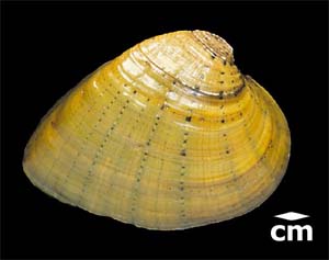
Illustration: Exterior photo of butterfly mussel (Ellipsaria lineolata).
What
Do Mussels Look Like?
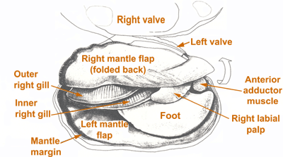
Illustration:
Drawing of mussel
soft tissues (after Burch 1973:7)
Inner
Bodies
Mussels have soft inner bodies and hard outer shells. The soft tissues
include a large muscular foot used for locomotion, an enveloping mantle
that secretes the shell, anterior and posterior adductor muscles that
enable to the animal to close its shells, labial palps that move food
particles to the mouth, and two pairs of gills. The gills have three
functions: (1) respirationlike fish, mussels use their gills to breathe,
(2) filter feedingthe gills move food particles to the mouth, and (3)
in females, the gills incubate baby mussels (larvae) until they are
mature and ready to be released.
Internal
organ systems include an open circulatory system powered by a heart;
a digestive system that consists of mouth, stomach, gut, and anus; a
decentralized nervous system that controls movement of the foot and
adductor muscles; and reproductive organs that usually occur separately
in male and female mussels.
 The
Shells
The
Shells
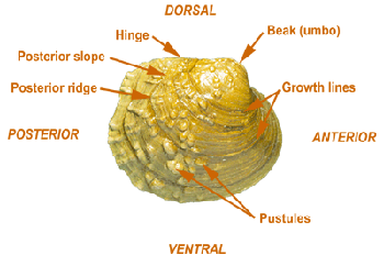
Each mussel has two shellsone left valve and one right valvethat protect
the soft-bodied animal from predators. The shells of different species
vary in size, shape, thickness, and color. Shells also vary in the presence
or absence of sculpturing (ridges or bumps) on the outer surface. Some
mussels have interlocking hinge "teeth" (pseudocardinal teeth and lateral
teeth) on the inside of the shell to help keep the two valves in proper
alignment. Other species are toothless. 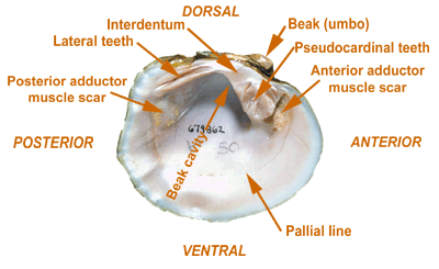
Illustration:
Exterior and interior photos of Purple wartyback (Cyclonaias tuberculata)
shell with parts labeled.
The
shells of mussels have three different layers. The outer layer (called
the periostracum) is made of organic material that may be yellow,
green, brown, or black. The middle layer (prismatic layer) is
made of elongate crystals of calcium carbonate (CaCO3). The
lustrous inner layer (nacre or mother-of-pearl layer) is made of plate-like
crystals of calcium carbonate and may be white, pink, salmon, or purple.
Look at the variety of shell colors in the mussel species and individuals
in the Photo
Gallery.
Related
Activity:
Mussel Anatomy (html) (pdf)
Mussel Identification (html)
(pdf) (interactive)